- Jun 04,2023
- Posted by: Savion
- Category:
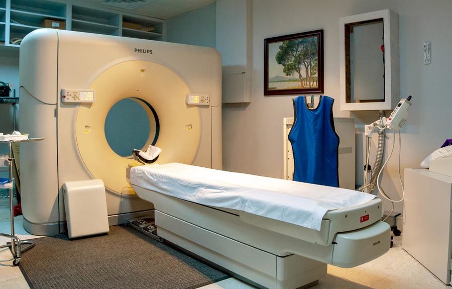
The axial tomography, generally known as CT, is similar to conventional imaging using radiation in the human body to create an image. By capturing sectional images, the sections are processed to generate three-dimensional images.
These images can be viewed on a computer screen, recorded on film, or converted to CD or DVD.
Axial tomography provides detailed and clearer images of internal organs such as bones, soft tissues, and vessels compared to regular radiation used for imaging tissues and vessels.
Radiology technicians form a specialized team to perform full-body scans using axial tomography and interpret the sections, facilitating early diagnosis of many tumors.
It is useful in cases such as motorcycle accidents and is used for acute chest and abdominal pain and acute breathing difficulties.
It is the best method for diagnosing many chest, abdominal, pelvic, liver, lung, pancreatic, and kidney tumors, as it confirms the presence of the tumor, its anatomical location, its relationship with adjacent organs, its size, and its degree of spread.
It is also crucial for screening, diagnosis, and treatment of conditions such as cerebrovascular diseases, renal failure, and vascular diseases that can cause human death.
It is an important means of diagnosing pulmonary embolism and aneurysms in the abdomen.
In many aspects, axial tomography resembles traditional simple imaging.
During CT scanning, the amount of radiation absorbed in the body is estimated by the radiation detectors rotating around the body.
Sometimes, during imaging, the device table rotates, and the rays are directed spirally.
The captured sections are collected and processed by the computer to create detailed images of internal organs in the body.
Thanks to advancements in sensor technology, multiple sections can be obtained during a single imaging procedure.
Multi-slice CT allows us to obtain numerous thin sections in a short period, providing precise details of internal organs. Modern CT scanners can perform scans of the body, especially in children, in a short time. This speed is crucial, especially for pediatric and geriatric patients and critical medical conditions. However, maintaining stillness during the imaging procedure remains essential.
For children, the aim is to reduce radiation exposure, and the dosage is calibrated according to the area being imaged and the section size. Contrast agents may be used to enhance image clarity.
During the imaging process, the patient may be asked to hold their breath.
Breathing and body movement can cause image distortion.
Generally, CT scanning is a painless, easy, and fast procedure. Multi-slice CT significantly reduces the imaging time for patients.
Unless under special circumstances, the patient is required to stay alone in the imaging room during the examination. Sometimes, during pediatric imaging, one of the parents is allowed to stay in the imaging room with the child after wearing a lead shield to protect against radiation. Communication between the technician, patient, and accompanying person inside the room during the imaging process is done through an internal microphone.
After completing the imaging procedure, the patient can return to their normal activities.